
Sunridge Medical patient education ·
Medically reviewed by Dr. Gioacchino Franco, NMD ·
Start with the impression, then connect it to the reason for the scan
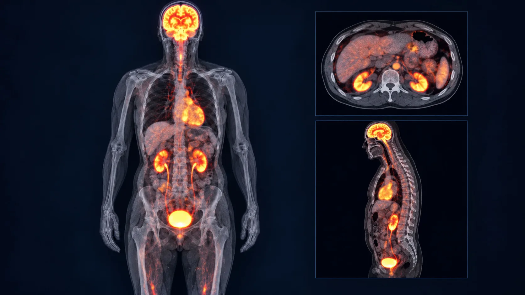
A PET/CT report is the interpreting physician’s written explanation of two kinds of information: PET shows where a radiotracer accumulates, while CT shows the anatomy of that area. The impression at the end summarizes the findings judged most important. It should be read together with the indication, comparison study, tracer, technique and detailed findings. 1, 2
The report can support staging, treatment planning or response assessment, but it is not a diagnosis by itself. The radiologist or nuclear-medicine physician integrates the uptake pattern with CT appearance, location, size, prior studies and the clinical history. Pathology may still be needed to establish what a finding represents.
PET shows tracer activity; CT shows structure
PET is molecular imaging. A radiotracer is designed to participate in or attach to a biological process. The scanner detects where that tracer accumulates. CT provides anatomic detail so the area of uptake can be located in a lymph node, organ, bone or other structure. 2, 3
The most common oncology tracer is fluorodeoxyglucose, or FDG, a glucose-like molecule. Many cancers use glucose actively and may accumulate FDG, but the tracer also appears in normal organs and in infection, inflammation and healing. Some cancers have low FDG uptake. 1, 6
Before interpreting a word such as “avid,” confirm which tracer was used. An FDG SUV cannot simply be compared with a PSMA or DOTATATE measurement.
“Whole-body” and “full-body” do not always describe the same scan range
In oncology PET/CT, “whole-body” commonly refers to a standard acquisition from the base of the skull or top of the head through the upper thighs. It does not automatically mean that every area from the top of the head through the toes was imaged. A true head-to-toe acquisition may be used for selected cancers, symptoms or clinical questions. Protocols vary by tracer, cancer type and imaging center. 1, 4
“Full-body” is often used conversationally and is not precise enough to identify the actual field of view. The report’s technique section should describe the imaged range. If the area you are concerned about is outside that range, ask the ordering clinician or imaging center whether it was included and whether another study is needed.
Read the report in this order
- Indication. Why the scan was ordered—initial staging, suspected recurrence, treatment response or another question.
- Tracer and technique. The radiotracer, dose, uptake time, scan range, blood glucose when relevant, and whether CT contrast was used.
- Comparison. The prior PET/CT, CT or MRI used to judge change. Confirm the comparison date.
- Findings. Observations organized by body region, including the site, size, uptake and CT appearance of important abnormalities.
- Impression. The interpreting physician’s synthesis and recommended correlation or follow-up.
When a finding appears in the body of the report but not the impression, ask whether it is considered incidental or clinically unimportant. When the impression recommends another study, biopsy or short-interval follow-up, ask which clinician will order it and on what timeline. 5
Translate common PET/CT language into questions
SUV measures relative tracer concentration—not a universal cancer score
SUV means standardized uptake value. It is a semiquantitative estimate of how much radiotracer is present in a selected area after accounting for factors such as administered activity and body size. SUVmax is the highest measured voxel value in the selected region. SUVmean averages the selected region. SUL is normalized to lean body mass.
There is no single SUV cutoff that reliably separates cancer from benign tissue. Infection and inflammation can have substantial FDG uptake, while some small, slow-growing or biologically different cancers can have modest uptake. The uptake pattern, CT appearance, location, clinical situation and pathology matter more than a standalone number. 4, 6
SUV also changes with preparation and technique: blood glucose and insulin, body size, time between injection and imaging, scanner calibration, reconstruction method, lesion size and how the region was drawn can all affect it. Serial SUVs are most useful when studies were performed in a comparable way. 4, 5
An area can “light up” for reasons other than cancer
FDG accumulates in cells using glucose actively. That includes many tumor cells, but also activated immune cells. Infection, inflammation, recent surgery, radiation change, healing tissue, fractures and some vaccinations can produce uptake. Normal brain, heart, kidney, bladder and bowel activity can be prominent. Muscle activity and brown fat can also affect the pattern. 4, 6
Recent biopsy, surgery, radiation, chemotherapy, growth-factor injection or vaccination can change uptake.
Very small lesions may be below PET resolution or have an underestimated SUV.
Not every tumor is strongly FDG-avid. A negative FDG study does not exclude all cancer.
The CT portion helps determine what structure corresponds to the uptake.
Treatment-response language depends on both metabolism and anatomy
A response report may describe decreased, increased, resolved, stable or new uptake. It may also describe changes in lesion size. A falling SUV can support metabolic response, but the significance depends on the cancer, treatment, timing, imaging protocol and the rest of the scan.
Some settings use formal response systems. Deauville scoring compares lymphoma uptake with reference tissues on a five-point scale. PERCIST is a structured metabolic response method used in selected settings. RECIST is primarily based on anatomic measurements. Do not translate one system into another without knowing which criteria the report used. 7
- Were the same tracer and similar protocol used?
- Which prior scan is the comparison?
- Did uptake change, size change, or both?
- Are there any new lesions?
- Could recent treatment or inflammation affect timing?
- Does the impression call the change response, stability or progression?
PET scan instructions: follow the imaging center’s protocol exactly
Preparation depends on the tracer, body region, indication, diabetes plan and whether the CT portion uses contrast. The appointment instructions from the imaging center override a general web guide. Call before the scan if the instructions are missing or conflict with your medication or diabetes plan.
Typical FDG preparation aims to reduce competing blood glucose and insulin effects and avoid uptake in exercising muscles. RadiologyInfo and procedure guidelines describe several hours of fasting, water for hydration, special diabetes instructions and attention to recent illness and treatment. 1, 4
What usually happens during and after a PET/CT scan
An IV is placed and the radiotracer is injected. For a typical FDG examination, you then rest quietly during an uptake period, often about an hour. Talking, chewing and muscle activity may be limited. During imaging, the table moves through the scanner and you must remain still. The scan itself may take roughly 15–60 minutes depending on the protocol. 1, 2
Afterward, most patients can resume normal activities unless the center gives different instructions. Hydration and frequent urination help clear tracer from the body. Follow the center’s instructions about close contact with infants or pregnant people, breastfeeding, contrast material and any sedating medication. 1, 8
Side effects are uncommon, but the tracer and CT contrast are different exposures
The radiotracer injection may cause a brief needle sensation, cold feeling, slight pain or redness. Significant reactions to diagnostic radiotracers are rare and are usually mild. Remaining still can be uncomfortable, and people with claustrophobia may feel anxious. 1
PET/CT uses ionizing radiation from the radiotracer and the CT portion. The diagnostic benefit is weighed against the relatively low radiation risk. Pregnancy and breastfeeding require advance discussion because the protocol or timing may need to change. 1, 8
Some PET/CT examinations include iodinated CT contrast; others do not. Contrast adds a separate set of considerations, including a prior contrast reaction, kidney function and other medical factors. Ask whether your CT is low-dose for localization or a diagnostic CT with contrast. 1, 9
- Possible pregnancy or breastfeeding
- Diabetes or difficulty fasting
- Kidney disease
- Prior contrast or radiotracer reaction
- Claustrophobia or inability to lie flat
- Recent surgery, infection, radiation, chemotherapy, vaccination or growth-factor medicine
- Breathing difficulty, facial or throat swelling, faintness or rapidly spreading hives
- Severe or worsening pain, swelling or redness at the IV site
- Any symptom your discharge instructions identify as urgent
A fictional example: how to work through the wording
Indication: Restaging after systemic therapy.
Comparison: PET/CT from three months earlier.
Findings: Left hilar lymph node now measures 0.9 cm with SUVmax 3.1, previously 1.6 cm with SUVmax 8.4. Stable 0.6 cm right upper-lobe nodule with mild uptake, SUVmax 2.8, indeterminate. No new FDG-avid lesion.
Impression: Interval decrease in size and metabolic activity of the left hilar node, compatible with treatment response. Stable indeterminate right upper-lobe nodule; continued attention on follow-up recommended.
This example is fictional and intentionally simplified. A real report can only be interpreted with the actual images, cancer type, treatment history and clinical context.
Bring these questions to the clinician reviewing your PET/CT
- Which tracer was used, and what biological activity does it measure?
- What was the scan intended to answer?
- Which prior study was used for comparison?
- Which findings are clearly important, and which are incidental?
- Does “uptake” here mean suspected cancer, inflammation, normal activity or uncertainty?
- Did the lesion change in uptake, size or both?
- Does this change my stage or treatment plan?
- Could recent treatment, infection, surgery or vaccination affect the finding?
- Is another scan, biopsy or laboratory test needed?
- What happens next, who orders it and on what timeline?
Keep the report in your cancer record and add a one-sentence summary after the appointment. Return to the Cancer Navigation Guide to organize the rest of the decision.
Common questions
Does a high SUV mean cancer?
No. A high SUV can occur in cancer, but also in infection, inflammation, healing and normal tissues. There is no universal SUV cutoff that proves whether a finding is malignant.
Is a lower SUV always good?
Not by itself. A lower SUV may support response when the same lesion is compared under similar conditions, but scanner technique, blood glucose, uptake time, lesion size and tumor biology can affect the value.
What does “no FDG-avid disease” mean?
It means the scan did not identify disease with abnormal FDG uptake. It does not exclude microscopic disease, very small lesions or cancers that are not strongly FDG-avid.
Can inflammation light up on a PET scan?
Yes. Activated immune cells use glucose and can accumulate FDG. Infection, surgery, radiation, healing tissue, fractures and some vaccinations can produce uptake.
Can I eat before a PET scan?
For many FDG PET/CT studies, patients are instructed to fast for several hours and drink only plain water. Other tracers and medical situations may have different instructions. Follow the imaging center’s protocol.
What if I have diabetes?
Call the imaging center for a specific plan for meals, insulin, oral medicines and appointment timing. Do not independently skip or change diabetes medication.
Are PET scan side effects common?
Significant radiotracer side effects are uncommon. Brief IV discomfort, a cold sensation, mild redness, anxiety or discomfort from lying still can occur. CT contrast, if used, has separate risks.
Can I be around family after the scan?
The center will give instructions based on the tracer and dose. Follow its guidance about close contact with infants or pregnant people and about breastfeeding.
Is a whole-body PET scan the same as a full-body PET scan?
Not necessarily. The terms are sometimes used loosely. A standard oncology whole-body PET/CT may cover the skull base or head through the thighs rather than the head through the toes. Check the technique section for the actual scan range and ask whether it matched the clinical question.
Research and sources
These sources support the medical and imaging information in this patient guide. Your own imaging center and care team remain the source for instructions and interpretation specific to you.
- RadiologyInfo.org (ACR/RSNA): PET/CT
- National Cancer Institute: Cancer imaging basics
- National Cancer Institute: CT scans and combined PET/CT
- EANM/SNMMI: FDG PET/CT procedure guideline for tumor imaging
- Society of Nuclear Medicine and Molecular Imaging: Essential elements of FDG PET/CT reporting
- Review: Infection and inflammation in oncologic FDG PET/CT
- National Cancer Institute: Imaging response criteria
- RadiologyInfo.org: Radiation safety
- RadiologyInfo.org: Contrast-material safety
Educational information only. This guide does not diagnose cancer, interpret an individual scan, or replace advice from the radiologist, nuclear-medicine physician or oncology team.