


A cancer diagnosis can arrive as a single sentence that changes the way the next few weeks feel: “Your cancer is Stage II.” You may hear the words, nod, and then realize you're still trying to understand what the number means. Does it describe the tumor itself, the chance of recovery, the treatment you'll need, or all three?
Cancer diagnosis stages provide a structured way for clinicians to describe how far a cancer has grown or spread. They help doctors communicate, compare treatment options, estimate prognosis, and coordinate care, but a stage is not a verdict or a complete description of you. Tumor biology, grade, biomarkers, overall health, and response to treatment also influence medical decisions.
The sections below translate the staging language into practical terms, including why stages differ between cancer types, how a stage is assigned, why it can change after surgery, and where an integrative evaluation may fit alongside conventional oncology. If you've recently been diagnosed, the newly diagnosed cancer resource from Sunridge Medical can also help you organize the questions that come up early.
Table of Contents
- The Moment You Hear Your Stage
- What Stages 0 Through IV Actually Mean
- The Major Staging Systems and Why They Differ
- How Doctors Determine Your Stage
- How Stage Shapes Prognosis and Treatment Choices
- Common Misconceptions Patients Bring to the Diagnosis
- Where Integrative Evaluation Fits and What to Do Next
The Moment You Hear Your Stage
The consultation room is quiet, but the language isn't. A physician explains the biopsy, mentions imaging, and then says, “The findings are consistent with Stage II breast cancer.” The patient hears “Stage II,” but the rest of the explanation turns into white noise. A spouse or parent sits nearby, silent, perhaps taking notes because the patient can't yet decide what to ask.
The first question is often about survival. The next questions usually arrive later, in the car or at home: How did they determine the stage? Could it change? What treatment comes next? Does Stage II mean the same thing for everyone?
Those questions are reasonable because a stage carries several meanings at once. It describes the known anatomic extent of disease, gives the oncology team a shared framework for planning, and helps clinicians discuss likely outcomes. It doesn't predict one person's future with perfect precision.
A stage is a clinical language for describing risk and extent. It isn't a sentence.
Consider a patient whose imaging shows a breast tumor without obvious distant spread. That patient may initially receive a clinical stage based on examination, imaging, and biopsy. If surgery later examines the tumor and nearby lymph nodes, the medical team may refine the stage using information that wasn't available before treatment.
You'll want clear answers to four core questions:
- What does my stage describe? Ask whether it reflects tumor size, lymph nodes, distant spread, or a combination.
- How was it determined? Request an explanation of the biopsy, scans, pathology, and any surgical findings.
- What treatment follows? Stage helps guide treatment, but receptor status, grade, and other biomarkers may change the plan.
- What does it mean for prognosis? Ask for a cancer-specific explanation rather than comparing your stage with someone else's.
The purpose of staging is to make a complex diagnosis more understandable to the care team and, eventually, to you. The number matters, but the details behind it matter more.
What Stages 0 Through IV Actually Mean
The Roman numerals offer a broad summary, while the TNM system supplies the details underneath. T describes the primary tumor's size or local extent, N describes regional lymph-node involvement, and M describes distant metastasis. This framework, developed by Pierre Denoix between 1943 and 1952, was adopted internationally in 1953 and became a major foundation of modern cancer staging through the work of the Union for International Cancer Control (UICC history of TNM).
Use a breast cancer example to see how the broad groups work. The exact tumor sizes and node combinations that qualify for each stage depend on the cancer type and its staging edition, so the descriptions below are intentionally general.
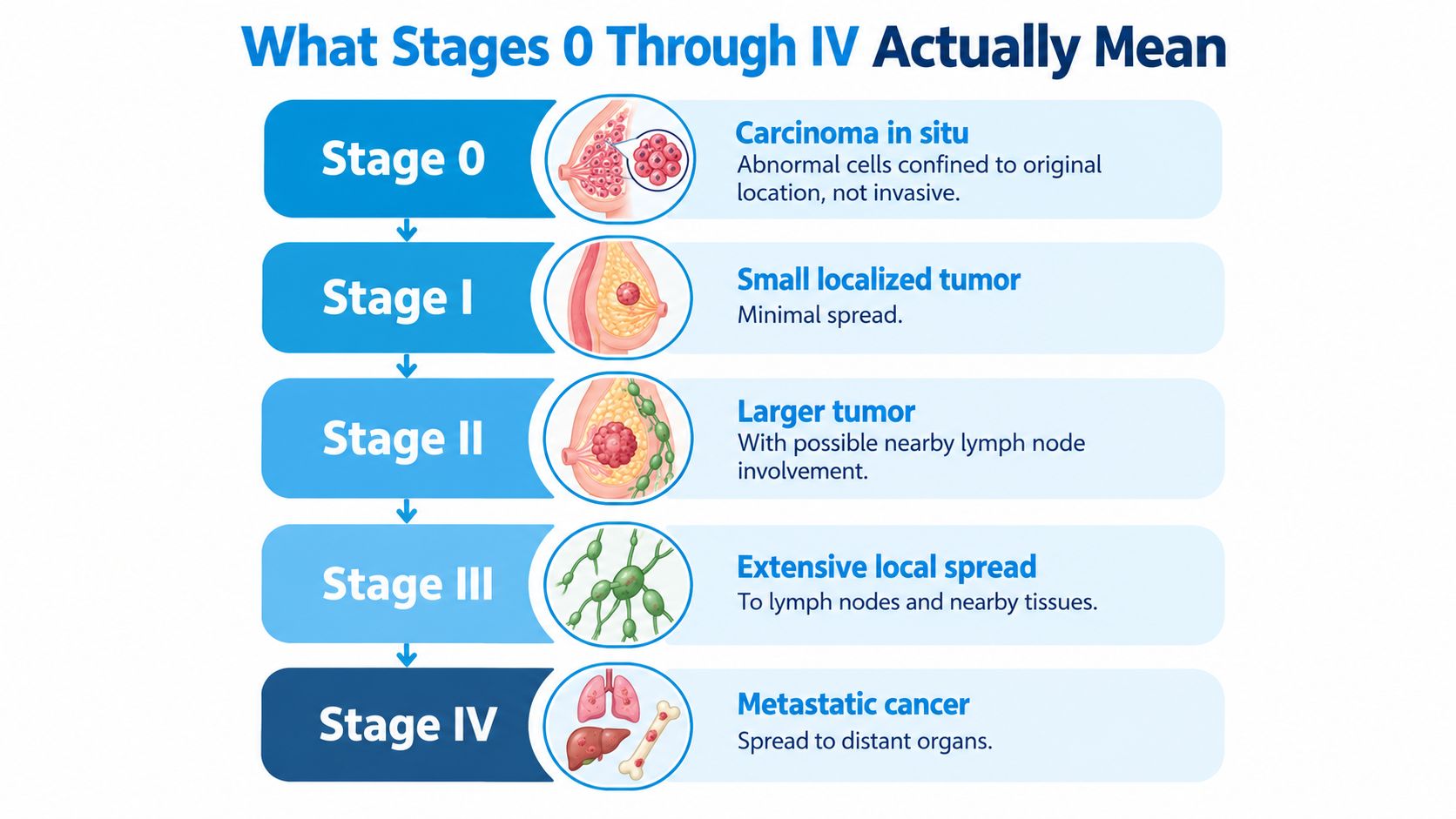
Stage 0
Stage 0 generally refers to carcinoma in situ. Abnormal cells remain confined to the location where they began and haven't invaded surrounding tissue. In a breast example, abnormal cells may be limited to a duct rather than growing into nearby breast tissue.
There's no distant metastatic disease in a Stage 0 definition. Because the cells haven't invaded, the treatment discussion often focuses on preventing progression and removing or controlling the abnormal area.
Stage I
Stage I usually describes a relatively small invasive tumor confined to the primary organ, with no regional lymph-node involvement in the broadest grouping. A small breast tumor found on imaging and confirmed by biopsy may fit this general pattern.
The word “small” isn't a universal measurement. Doctors use cancer-specific rules that combine tumor dimensions, local invasion, node findings, and sometimes biological features.
Stage II
Stage II generally indicates a larger tumor, more local extension, or limited involvement of nearby lymph nodes, while distant metastasis hasn't been identified. A breast tumor might be larger than a Stage I tumor, or a smaller tumor might have selected regional node involvement.
That doesn't automatically mean the cancer has become untreatable. It means the team must account for a greater local or regional burden when planning surgery, radiation, systemic therapy, or combinations of these approaches.
Stage III
Stage III usually reflects more extensive local or regional disease. Several lymph nodes may be involved, or the tumor may have grown into nearby structures, but the defining pattern remains the absence of known distant metastasis.
The distinction between regional and distant spread is clinically important. A lymph node near the original organ is different from cancer found in a distant liver, lung, bone, or other organ.
Stage IV
Stage IV means distant metastatic disease has been identified. The primary tumor may be small or large, and regional nodes may or may not be involved, but the presence of distant spread places the cancer in a different treatment category.
A Roman numeral doesn't carry the same meaning across diseases. Stage II breast cancer, Stage II colon cancer, and Stage II pancreatic cancer use different criteria and can have different treatment pathways. Your oncologist should explain the cancer-specific staging rules rather than relying on the numeral alone.
The Major Staging Systems and Why They Differ
Cancer staging systems differ because cancers behave differently and arise in different anatomic settings. A system that accurately describes a solid tumor may not capture the biology or distribution pattern of lymphoma, a gynecologic cancer, or thyroid cancer.
The TNM system is the major framework for many solid tumors. T may include numerical and letter modifiers that describe size, depth, or invasion. N identifies whether regional nodes contain cancer and, depending on the cancer, how many or which nodes are involved. M records whether distant metastasis is present. Clinicians then combine these findings into a stage group, often expressed as Stage 0 through Stage IV (clinical and pathologic TNM overview).
Other systems use different organizing principles:
- Ann Arbor staging is used for lymphomas and considers the distribution of involved lymph-node regions and whether systemic “B symptoms” are present.
- FIGO staging is used for gynecologic cancers and emphasizes the local and regional pattern relevant to organs such as the cervix, uterus, ovary, or vulva.
- RAI staging is associated with thyroid cancer and incorporates disease features specific to thyroid malignancy and its spread.
| Staging System | Cancers It Applies To | Primary Criteria |
|---|---|---|
| TNM | Many solid tumors | Primary tumor extent, regional nodes, distant metastasis |
| Ann Arbor | Lymphomas | Nodal regions, extranodal involvement, systemic symptoms |
| FIGO | Gynecologic cancers | Cancer-specific local, regional, and distant spread |
| RAI | Thyroid cancer | Thyroid cancer features, age-related considerations, and spread |
Roman numeral groups make conversations easier, but they compress detailed findings. Two cancers can both be called Stage III while differing in tumor biology, treatment sensitivity, and expected course. That's why a Stage III colon cancer shouldn't be compared directly with a Stage III lymphoma without knowing the disease-specific system.
The staging process also includes unknown when clinicians don't yet have enough information. The American Cancer Society explains that stage depends on tumor size, location, lymph nodes, and distant spread, and that further testing can refine the initial picture (cancer staging overview).
How Doctors Determine Your Stage
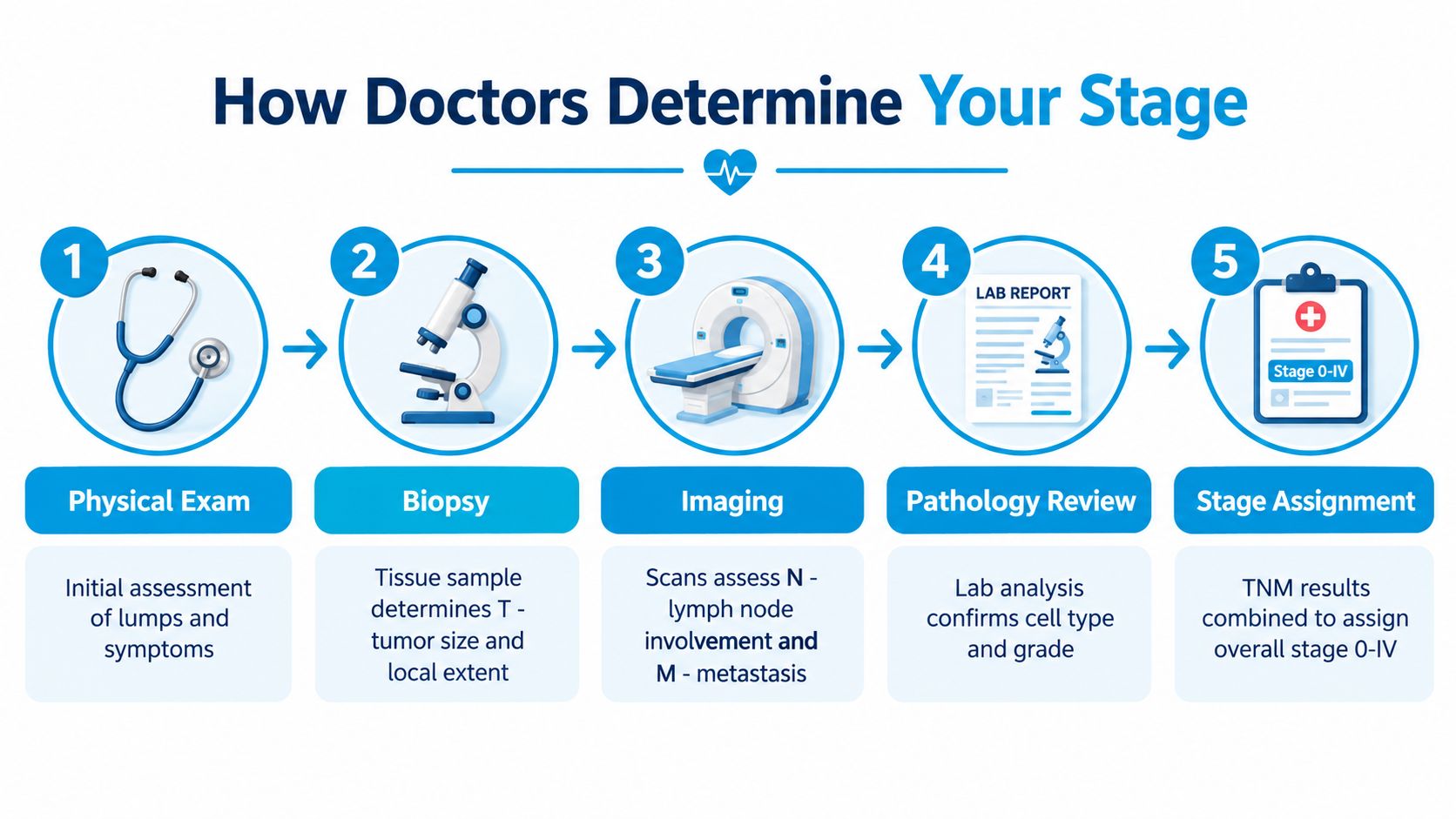
Staging usually develops through several encounters rather than one test. A patient with a new breast lump may first have a physical examination, followed by diagnostic imaging and a biopsy. Each step answers a different question.
The initial findings
The physical examination helps the clinician assess a lump, skin changes, regional nodes, or other signs of local disease. Imaging, such as ultrasound, mammography, CT, MRI, or PET, can show the tumor's location and help evaluate lymph nodes or possible distant lesions.
A biopsy confirms what the abnormal tissue is. Pathologists examine the tissue to identify the cancer type, assess grade, and perform relevant biomarker testing. In breast cancer, hormone receptor and HER2 testing may influence treatment even though those results aren't interchangeable with an anatomic stage.
Clinical and pathologic staging
Doctors assign a clinical TNM stage before treatment using the examination, imaging, biopsy, and available laboratory information. If surgery removes the tumor or samples lymph nodes, a pathologist can examine those tissues directly and assign a pathologic TNM stage afterward.
That distinction explains why a stage may change. Imaging might suggest that a lymph node is clear, but surgical pathology may find microscopic cancer. Conversely, treatment before surgery may shrink the tumor and leave less viable cancer in the specimen, leading to a post-treatment stage that reflects treatment response.
Ask which stage you're being given: clinical, pathologic, or post-treatment. The label provides important context.
A multidisciplinary tumor board may review the findings with medical oncology, surgery, radiation oncology, radiology, and pathology. For patients who want to understand the diagnostic workflow and available testing, Sunridge Medical's cancer diagnostic testing information offers another patient-oriented explanation. Laboratories also rely on specialized environments, and readers interested in technical standards can review SEFA-compliant pathology furniture as an example of how pathology workspaces are designed around laboratory requirements.
For a visual explanation of the process, this video provides additional context:
How Stage Shapes Prognosis and Treatment Choices
Stage strongly influences prognosis because the anatomic extent of cancer often reflects how difficult it is to remove or control. Population data from England illustrate the difference between early and late diagnosis. For breast cancer, 1-year survival is about 97% at Stage 1 and about 67% at Stage 4, while colorectal cancer falls from about 90% at Stage 1 to about 46% at Stage 4 (Office for National Statistics cancer survival estimates).
Those figures describe groups, not an individual prediction. Tumor grade, receptor status, molecular profile, age, other health conditions, treatment access, and response all modify the outlook. In the same dataset, melanoma had the highest reported 1-year net survival among the listed cancers, at 97.4% for men and 98.6% for women, while pancreatic cancer had the lowest, at 23.7% for men and 25.3% for women (same ONS dataset).
Treatment intent changes with extent
Early disease may be approached with local treatment intended to eliminate the cancer, such as surgery or radiation. Stage II and Stage III disease often require a combination of local and systemic treatments, depending on cancer type and biomarkers. Stage IV treatment frequently emphasizes controlling disease, preserving function, reducing symptoms, and maintaining quality of life, although some metastatic cancers can be treated aggressively in selected circumstances.
Colon cancer provides a stark example of the stage gradient. AJCC-based analyses report 5-year survival of about 93.2% for Stage I colon cancer and about 8.1% for Stage IV, with outcomes falling through intermediate stages as nodal burden rises or distant metastasis appears (AJCC-based colon cancer survival analysis). The transition from N0/M0 disease to M1 disease is a major prognostic and treatment inflection point.
| Stage Group | Typical Prognosis Range | Treatment Intent | Common Modalities |
|---|---|---|---|
| Stage 0 | Often highly favorable when fully treated | Prevent invasion or recurrence | Local excision, surgery, selected radiation |
| Stage I | Often favorable, cancer-specific | Curative when appropriate | Surgery, radiation, selected systemic therapy |
| Stage II | Variable and cancer-specific | Frequently curative | Surgery with chemotherapy or radiation when indicated |
| Stage III | Variable, with greater regional risk | Curative or disease-controlling | Combined local and systemic treatment |
| Stage IV | Broadly variable | Control, symptom relief, and quality of life, with selected intensive approaches | Systemic therapy, targeted therapy, immunotherapy, surgery or radiation in selected cases |
The treatment plan should connect the stage to the biology, not use the stage as a shortcut. Sunridge Medical's integrative cancer treatment by stage clinical reference guide discusses how an individualized plan can be considered alongside conventional oncology.
Common Misconceptions Patients Bring to the Diagnosis
A patient may leave the first oncology appointment believing that the stage is permanent, universal, and predictive of an exact future. None of those assumptions is reliable.
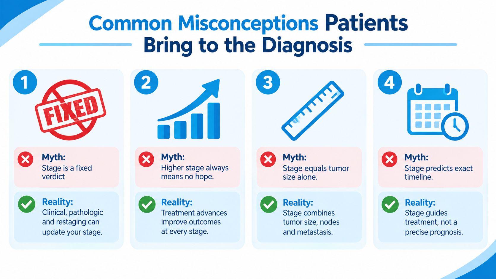
Misconception one: the stage can never change. A clinical stage is based on information available before treatment. Surgery may reveal lymph-node involvement that imaging didn't show, or a biopsy review may clarify the tumor type. If chemotherapy or another treatment occurs before surgery, the later pathology may describe how much cancer remains after treatment. The medical record may preserve the original clinical stage while adding a pathologic or post-treatment stage.
Misconception two: the same Roman numeral means the same severity. Stage I breast cancer and Stage I pancreatic cancer aren't interchangeable labels. Each disease uses criteria that reflect its anatomy, patterns of spread, and biology.
Stage isn't the whole diagnosis
Misconception three: stage equals tumor size. Stage combines the primary tumor, regional lymph nodes, and distant metastasis. A smaller tumor with distant spread may represent more advanced disease than a larger tumor confined to its original organ.
Misconception four: a higher stage always means there's no hope. A higher stage often means the cancer requires a more complex strategy, but treatment options vary substantially by cancer type and biology. Stage IV disease can sometimes be controlled for meaningful periods, and some patients receive treatment directed at specific molecular features.
The reverse misconception also causes harm. A lower stage doesn't mean the diagnosis can be ignored. Early-stage cancers still require timely treatment, surveillance, and attention to the features that influence recurrence risk.
Use stage as a working snapshot of known disease extent, then ask what the pathology and biomarkers add to that snapshot.
The most useful questions are concrete: “What is my clinical stage?” “Could surgery change it?” “What is my tumor grade?” “Which biomarkers affect my treatment?” and “What does this stage mean for this specific cancer?”
Where Integrative Evaluation Fits and What to Do Next
An integrative oncology evaluation doesn't replace TNM staging, tumor-board review, surgery, chemotherapy, radiation, or other established cancer treatment. It adds a broader assessment of the patient's functional context, including nutrition, sleep, symptoms, medication interactions, comorbidities, physical capacity, and treatment goals.
A careful clinic may review the pathology diagnosis, imaging, treatment history, medication list, nutritional status, and relevant laboratory data. Depending on the patient and the evidence, clinicians may also discuss inflammatory markers, metabolic health, immune-related findings, or gastrointestinal symptoms. These assessments may help identify supportive needs, but they don't change the anatomic stage or prove that an unestablished therapy can treat cancer.
A coordinated evaluation
The practical process often begins with records. Bring pathology reports, imaging reports, operative notes, medication and supplement lists, and the names of the oncology clinicians involved. A consultation can then distinguish established treatment from complementary support, off-label use, or investigational approaches.
Sunridge Medical describes an integrative oncology model that reviews the cancer diagnosis and conventional treatment context while considering individualized supportive and adjunctive options. Its role of integrative oncology in modern cancer care provides additional discussion of how this type of evaluation can be coordinated with oncology care.
Useful preparation includes:
- Request the records: Ask for pathology reports, imaging reports, treatment summaries, and, when appropriate, access to pathology slides for review.
- List everything you take: Include prescriptions, over-the-counter products, vitamins, herbs, and supplements because interactions can affect treatment safety.
- Clarify the purpose: Decide whether you want a second opinion, symptom support, help preparing for treatment, or a coordinated review of options.
- Keep clinicians informed: Tell your oncologist about every added therapy, infusion, supplement, or procedure before starting it.
Integrative care should remain collaborative. A patient seeking lifestyle support may also find it useful to explore reputable resources about related integrative topics, such as treating ADHD naturally, while recognizing that advice for another condition doesn't automatically apply during cancer treatment.
The most important shared language remains the stage, TNM findings, pathology, biomarkers, and treatment response. Your integrative clinician and oncology team should be working from the same records and communicating about safety.
Sunridge Medical in Scottsdale offers individualized integrative oncology evaluations that review cancer staging, pathology, imaging, treatment history, symptoms, and supportive care needs alongside conventional oncology. Visit Sunridge Medical to request a consultation and discuss whether a coordinated evaluation is appropriate for you or your family.