
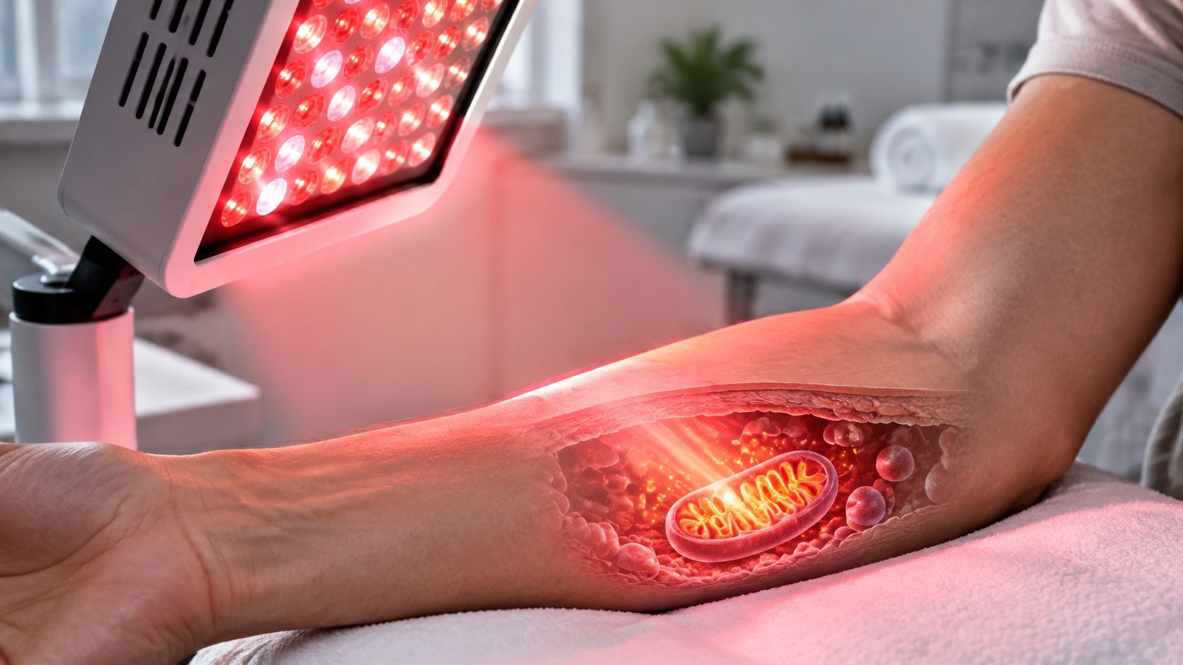
How does red light therapy work? Photobiomodulation uses red light, typically 600 to 700 nanometers, and near-infrared light, typically 780 to 1,100 nanometers. Mitochondria absorb light in these ranges, influencing cellular energy production and signaling without heating tissue. This proposed mechanism helps explain how light could affect cells, but it does not establish benefits for every device or condition.
Two products can both glow red yet differ in their wavelengths, doses, instructions, and supporting evidence. Wavelength and dose shape the biological response, while the condition being studied determines whether that response has demonstrated clinical value. Separating a plausible cellular mechanism from a proven patient benefit is key to understanding photobiomodulation.
Table of Contents
- How Red Light Therapy Works Inside Cells
- Wavelengths and Doses That Change the Response
- Where Evidence Is Stronger and Where It Is Not
- Safety Limits When More Light Is Not Better
- Red Light Therapy Beside Cancer or Regenerative Care
- What an Individualized Evaluation May Involve
- Decide Whether a Consultation Is Appropriate
How Red Light Therapy Works Inside Cells
A useful analogy is a dimmer that changes how a power plant runs. It isn't a furnace burning the building, and it isn't a laser cutting tissue away. The light is meant to influence how a cell produces and uses energy while leaving the tissue intact.
Say a clinician positions a red and near-infrared device over skin. Photons enter the tissue, some are absorbed, and some pass through or scatter. What matters is not that the area looks red. What matters is whether light at a usable wavelength reaches a target inside the cell at a dose that changes signaling.

The Proposed Sequence Inside Mitochondria
A review of photobiomodulation mechanisms describes non-thermal red light at typically about 600 to 700 nanometers and near-infrared light at commonly about 780 to 1,100 nanometers, with cytochrome c oxidase as the leading proposed photoacceptor. The sequence is usually explained in three steps:
- Photon absorption: Cytochrome c oxidase, the terminal enzyme in the mitochondrial electron-transport chain, absorbs photons at certain wavelengths.
- Nitric oxide release: Absorption may alter copper-containing centers in the enzyme and promote release of nitric oxide, which can otherwise compete with oxygen at the enzyme's active site.
- Energy signaling: With oxygen binding improved, electron transport, mitochondrial membrane potential, proton movement, and production of adenosine triphosphate (ATP), the cell's main energy currency, may increase.
A recent review of photobiomodulation biology places commonly used red wavelengths at approximately 620 to 700 nanometers, near-infrared treatments at generally about 700 to 1,100 nanometers, and frequently studied clinical wavelengths at 670, 740, and 850 nanometers. That range matters because absorption isn't flat. The 700 to 780 nanometer interval overlaps a relative absorption trough for cytochrome c oxidase, so two wavelengths that look similar to the eye may not interact with the enzyme in the same way.
Readers who want the older laser terminology placed beside the modern photobiomodulation framework can consult the Omega Lasers guide to LLLT, which provides background on low-level laser therapy as a related form of light-based care.
Why Effects Might Outlast the Exposure
Work associated with Tiina Karu introduced retrograde mitochondrial signaling to explain how a brief exposure might influence a cell for hours, days, or weeks. In plain terms, mitochondria don't only make ATP. They also send signals that can affect processes beyond the moment the light is switched off.
Reviews describe downstream changes involving oxygen consumption, redox signaling, growth-factor synthesis, oxidative stress, and inflammatory signaling. Those are biological findings, not promises about a diagnosis. A change in a signaling pathway in a laboratory or early clinical study doesn't establish that a home panel will change pain, healing, or disease course for a particular person.
A plausible mechanism explains why researchers study a treatment. It doesn't, by itself, prove that a specific device helps a specific condition.
The response is also dose-dependent. Wavelength, irradiance, exposure time, treatment frequency, tissue depth, and delivered energy density all influence what happens, so more light isn't automatically better. That single point explains much of the confusion around consumer claims. A device can deliver red light and still miss the wavelength, dose, or tissue target used in a study.
Wavelengths and Doses That Change the Response
Wavelength identifies the part of the light spectrum used; dose describes the exposure delivered to tissue. Treat color as the starting label and dose as the measurable exposure. The ranges below are reported in an Aesthetic Surgery Journal review of photobiomodulation parameters, except where the source is specified in the row.
| Parameter | Typical published values | What it changes |
|---|---|---|
| Visible red light | About 600 to 700 nm; skin studies often use 630 to 660 nm | Exposure in more superficial tissue |
| Near-infrared light | About 780 to 1,100 nm | Can reach deeper tissue than visible red, depending on tissue and exposure |
| Often studied wavelengths | 670, 740, and 850 nm | Examples of wavelengths used in research, not a prescription |
| Reduced cytochrome c oxidase | Absorption peaks near 620 nm and 710 to 790 nm | The enzyme absorbs light differently across these ranges |
| Oxidized cytochrome c oxidase | Absorption peaks near 650 to 680 nm and 820 to 830 nm | Absorption varies with the enzyme's oxidation state |
| Relative absorption trough | About 700 to 780 nm | Cytochrome c oxidase absorption is relatively weaker |
| Optical window | About 600 to 970 nm, within a broader 600 to 1,100 nm range | Water absorption rises above about 970 nm |
| Irradiance | About 5 mW/cm² to 5 W/cm² | Power delivered per unit area |
| Fluence | Joules per square centimeter, calculated from irradiance and exposure time | Total energy delivered to an area |
Wavelength, irradiance, and fluence describe different parts of an exposure. Wavelength, measured in nanometers, locates light on the spectrum. Irradiance measures power reaching each square centimeter; fluence measures accumulated energy per square centimeter. A device's settings and its distance from the skin affect how much energy reaches the treated area.
The review reports different absorption peaks for reduced and oxidized cytochrome c oxidase, a mitochondrial enzyme. Those laboratory measurements help explain why wavelength matters, but they do not identify a universally effective setting for a particular condition.
A 2025 review summarized in a PubMed record on photobiomodulation wavelengths found that 68.42% of the studies it examined used wavelengths from 630 to 890 nanometers. That figure describes the wavelengths used in the reviewed research, not a recommended dose for an individual.
Why Distance and Time Change the Exposure
Moving a panel farther from the skin generally reduces the power reaching each square centimeter. A shorter session also reduces total fluence. The device may stay the same, yet the delivered exposure changes.
For that reason, session length alone cannot define a dose. Patients comparing research parameters with everyday use may ask about how much red light therapy per day. A useful comparison also accounts for the device, distance, wavelength, irradiance, treated area, and exposure time. Published parameters are research examples, not a home dosing plan.
Reading rule: A stated color and session time do not describe the full exposure.
Tissue also changes how light travels. Water absorption rises above about 970 nanometers, and compounds such as flavins, hemoglobin, and melanin absorb some light. The wavelength that reaches a target therefore depends on both the light source and the tissue it passes through.
Where Evidence Is Stronger and Where It Is Not
What can red light therapy reasonably be expected to do? Early experiments helped direct research toward tissue repair and cellular signaling. Evidence still needs to be read by condition and outcome: a finding about wound healing, pain, or a treatment side effect does not establish a benefit for every related disease.
| Patient question | What research suggests | What it does not show |
|---|---|---|
| Can it affect tissue repair? | Early observations and later research have examined possible effects on tissue repair | That it heals every wound or works for every person |
| Can it affect pain or inflammation? | Research examines pain and inflammatory signaling | That it is an established treatment for every pain condition |
| Does it help arthritis? | A historical review of low-level light therapy describes systematic reviews that found evidence insufficient for some osteoarthritis and rheumatoid arthritis uses | That it should replace established arthritis care |
| Will it rejuvenate skin? | Some commercial studies have examined red-light exposure for cosmetic outcomes | That a cosmetic result treats disease |
| Can it support cancer care? | Research has examined selected side effects related to cancer treatment | That it treats cancer or replaces oncology care |
A review of the field describes photobiomodulation research across red and near-infrared wavelengths. The distinction matters: a research field can be well defined while a treatment for a named disease remains unproven. Findings for one condition, device, or outcome do not automatically apply to another.
Wound healing also depends on factors such as circulation, infection, pressure, nutrition, and diabetes control. Light is one possible part of care, not a full wound assessment. Readers considering wound care can review wound care treatment options from Comprehensive Wound Services to understand how clinical services address wounds beyond a single device.
Cosmetic and scalp claims need their own evidence because the target and measured outcome differ from those in wound or pain studies. For example, information about red light therapy on the scalp should be assessed against evidence for scalp and hair goals, rather than assumed to follow from skin-rejuvenation research.
Four Questions That Expose Weak Claims
- Which condition was studied? A result for a treatment side effect does not establish a result for the underlying disease.
- Was the research controlled and conducted in humans? Cell and animal findings can guide research, but they do not establish patient benefit.
- What wavelength and fluence were used? Without these parameters, it is difficult to compare the study with a particular device or session.
- What outcome changed? A symptom score, treatment side effect, appearance, and disease progression are different outcomes.
The most useful conclusion is specific: assess the condition, the light parameters, and the measured outcome together. Photobiomodulation may have a supportive role in selected settings, with care coordinated around the patient's broader treatment plan.
Safety Limits When More Light Is Not Better
Higher intensity and longer exposure don't automatically produce a stronger benefit. Photobiomodulation has a dose-dependent response, and excessive exposure can injure skin. That point is easy to miss because many sessions feel gentle, but comfort during exposure isn't proof that any dose is appropriate.
A report of two randomized dose-escalation trials of high-fluence LED red light found dose-limiting blistering or prolonged erythema at approximately 480 to 640 joules per square centimeter. Mild transient erythema and hyperpigmentation were also reported, and no serious adverse events occurred in those trials. Those numbers are a warning about very high fluence in a research setting. They aren't a target dose, and they don't guarantee that lower consumer exposures are risk-free for every person.
What a Clinician Should Review First
- Skin type and prior reactions: Pigmentation changes and prolonged redness don't affect every patient equally.
- Photosensitivity and medicines: A full medication review matters because some drugs increase light sensitivity.
- Eye protection: The treatment area, device position, and eye history determine what protection is appropriate.
- Treatment area and indication: Light over intact skin for a cosmetic goal raises different questions from light near a wound, a scar, or a cancer treatment field.
- Diagnosis certainty: An uncertain diagnosis should be clarified before a biologically active adjunct is added.
Extra caution is warranted with active malignancy, eye disease, photosensitizing medication use, or any plan to aim light at a tumor site. In those settings, the question belongs with the clinician directing care, and patients exploring coordinated cancer support can read about integrative oncology programs as context for how supportive options are reviewed beside conventional treatment.
Home devices may not match research equipment in wavelength accuracy, output, beam pattern, or distance. Published results therefore don't transfer automatically to every consumer product, even when marketing uses the same color name.
New severe pain, rapidly worsening skin injury, vision symptoms, or a suspected cancer complication need urgent or conventional medical assessment, not another light session.
No responsible safety discussion ends with a universal home schedule, distance, or minute count. The safe exposure is the one selected for a defined indication after the person's skin, eyes, medicines, diagnosis, and treatment area have been reviewed.
Red Light Therapy Beside Cancer or Regenerative Care
For someone receiving cancer treatment, the practical question is whether a specific light exposure could help with a particular treatment-related side effect without conflicting with the oncology plan. Photobiomodulation may have a supportive role for selected concerns, but it does not replace surgery, radiation, systemic therapy, or an oncologist's recommendations.
A 2020 oncology-focused review of photobiomodulation reported that available in-vivo studies and clinical trials found photobiomodulation safe in relation to tumor growth while supporting management of selected cancer-treatment-related complications. The review supports light for selected side effects, not for directing light over a tumor.
Wavelength, dose, location, timing, cancer type, treatment plan, and evidence quality all shape the question. The reported benefits concerned specific treatment-related side effects, not survival, disease reversal, or replacement of standard treatment. The distinction matters: a possible effect on symptoms does not establish an effect on the cancer itself.
Photobiomodulation also differs from light-activated drug treatment. In photodynamic therapy for cancer, a photosensitizing drug is combined with light. That is a different intervention from red light alone, with different risks and evidence.
Questions to Bring to the Oncology Team
- Target: Which treatment-related side effect is the proposed target?
- Location: Has the oncologist reviewed exposure near the tumor or treatment area?
- Timing: Could the proposed timing affect coordination with surgery, radiation, or systemic therapy?
- Uncertainty: Which parts of the plan have human evidence, and which remain uncertain?
Name the outcome first: walking distance, wound closure, sleep, or return to activity. Each goal needs its own way to track change. A broad claim about repair is difficult to assess unless it is tied to a specific function or symptom. Recovery care should be judged by whether that defined outcome changes, not by the general promise of cellular support.
What an Individualized Evaluation May Involve
An individualized evaluation starts with the diagnosis, not the device. The clinician reviews what's already known, who is directing oncology or other conventional care, and what outcome the patient hopes light might influence. Sometimes the conclusion is that photobiomodulation isn't relevant. Sometimes more records are needed, or the question should return first to the oncology or primary care team.
History That Changes the Decision
A complete medication list matters, including photosensitizing drugs, supplements, and recent changes made by other clinicians. Eye history, skin type, prior reactions to light, and any history of pigment change after procedures also shape the risk review. The area of concern needs direct attention because intact skin, a healing wound, a scar, and tissue near a cancer treatment field aren't equivalent targets.
Circulation can change both the goal and the urgency. A slow-healing leg wound, for example, shouldn't be attributed to low cellular energy until arterial disease and other causes have been considered. Readers who need background on that separate diagnostic issue can start with understanding peripheral artery disease before assuming a light-based approach addresses the underlying problem.
Patients comparing regenerative options face a similar sorting task. A discussion of platelet-rich plasma vs stem cell approaches, for instance, involves injections, tissue targets, and candidacy questions that don't transfer to a non-invasive light session. Keeping categories separate prevents one therapy's evidence from being borrowed by another.
If Light Is Considered and What to Bring
If light remains a reasonable question after review, the clinician compares published research parameters against the individual. That includes wavelength, irradiance, fluence, time, distance, and treated area, plus documentation of eye protection and skin monitoring. Home devices may not match research equipment, so a published result still doesn't transfer automatically to a product used outside a clinic.
- Recent records: Visit notes that state the current diagnosis and plan.
- Imaging or pathology reports: Reports that clarify the area or condition under discussion.
- Medication list: Prescriptions, over-the-counter products, and supplements with doses.
- Written questions: The target outcome, the evidence, and who else needs to approve.
Bring the goal in one sentence: A defined target, such as a named side effect or a specific area of skin, leads to a clearer evaluation than a general request for more energy.
Decide Whether a Consultation Is Appropriate
A useful decision starts with three questions: Can the reader explain photobiomodulation as non-heating light that may influence mitochondrial signaling? Do they understand that wavelength and fluence shape exposure, so color and minutes alone do not describe a treatment? Can they name what the evidence does and does not show?
Evidence applies to specific indications, devices, and outcomes. It does not establish disease reversal or a survival benefit. With those three points clear, a consultation becomes a focused clinical conversation.
Adults in Scottsdale, Phoenix, and the greater Phoenix area, as well as people who travel to Arizona for care, may contact Sunridge Medical to ask whether an evaluation, consultation, or second opinion is appropriate. Suitability depends on an individualized review of medical history, diagnosis, medicines, current clinicians, and goals. For a patient receiving oncology care, that review should include how any proposed supportive approach fits alongside the existing care plan.
Sunridge Medical offers Red Light Therapy as private, full-body photobiomodulation sessions after staff setup and instructions. A consultation can help clarify whether that option is relevant to the concern under discussion and what questions need to be addressed with the patient's other clinicians.
Before contacting a clinic, gather current records, a medication list, and a one-sentence description of the goal. A specific concern gives the clinician a clearer starting point for evaluating whether light therapy is appropriate.